

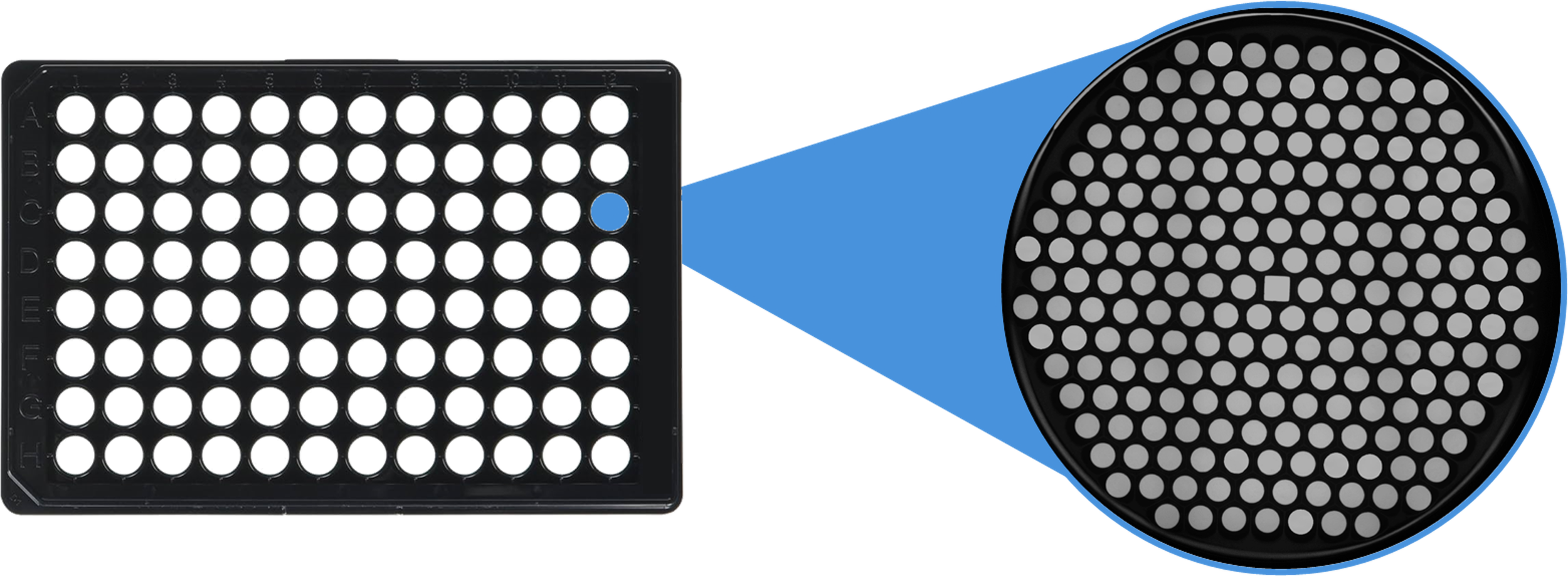
Nanowell Plates
for imaging single-cells, organoids and model-organisms
Nanowell plates
ImageCyte makes nanowell imaging plates that arrange biological samples into tiny, defined spaces while staying compatible with standard lab workflows.
Better data, higher confidence.
Explore our applications
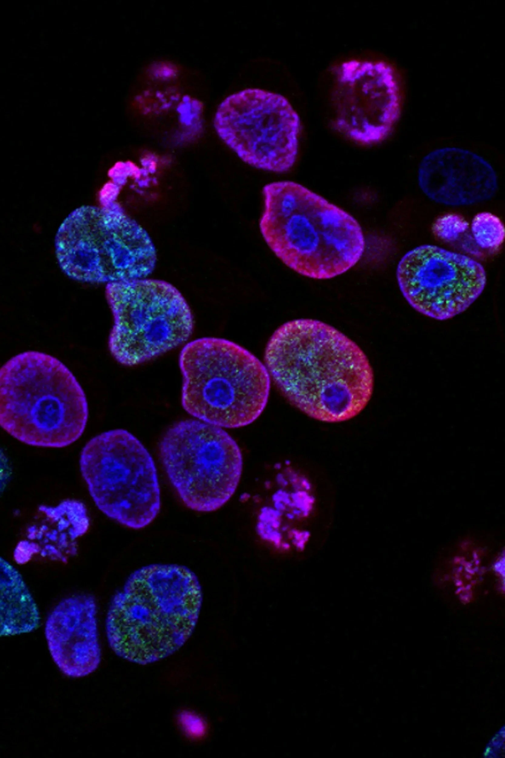
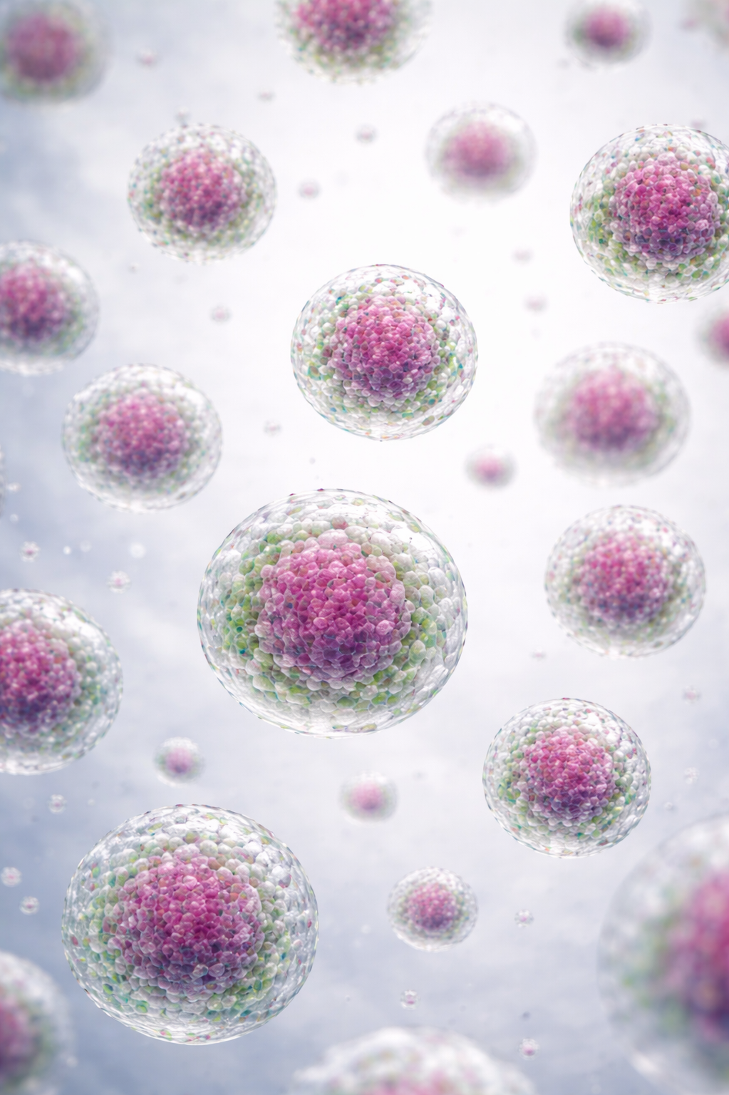
Single-cell Isolation
Multi-cell Observations
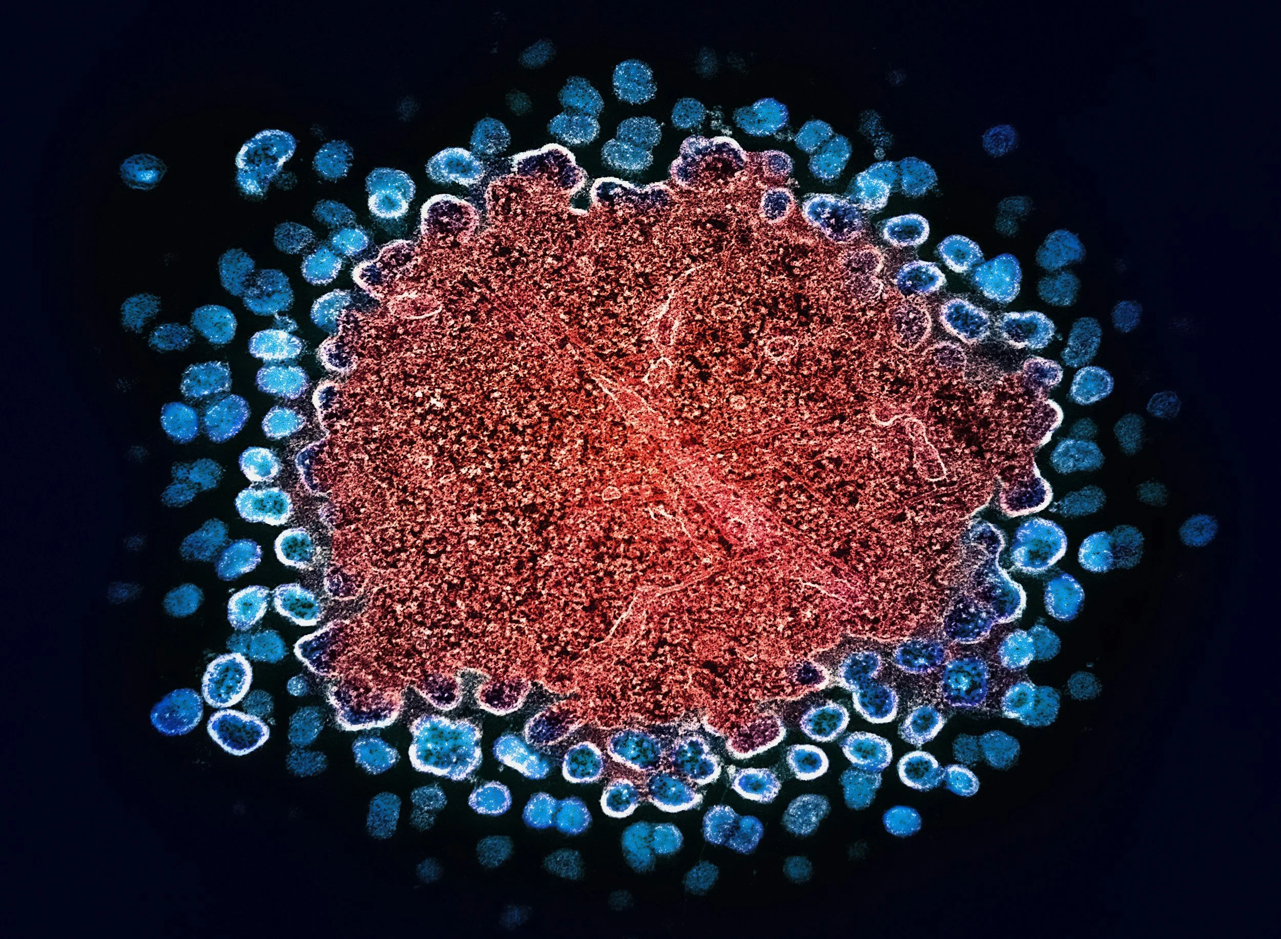
Organoids
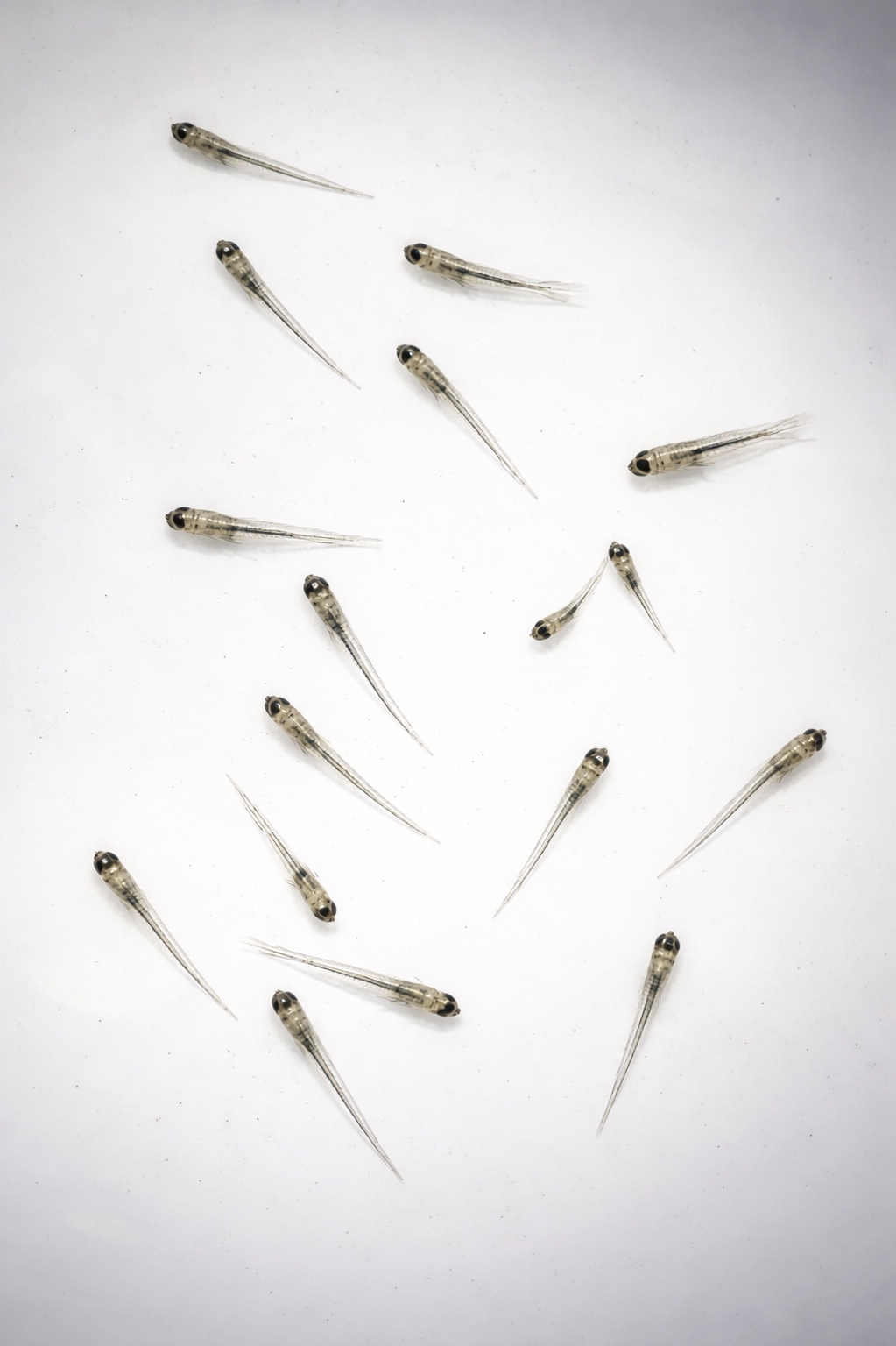
Zebrafish
Interested in evaluating ImageCyte plates for your workflow?
We work directly with research groups to assess compatibility, imaging performance, and throughput for specific applications.
Request an evaluation
or
Contact us to discuss your experiment